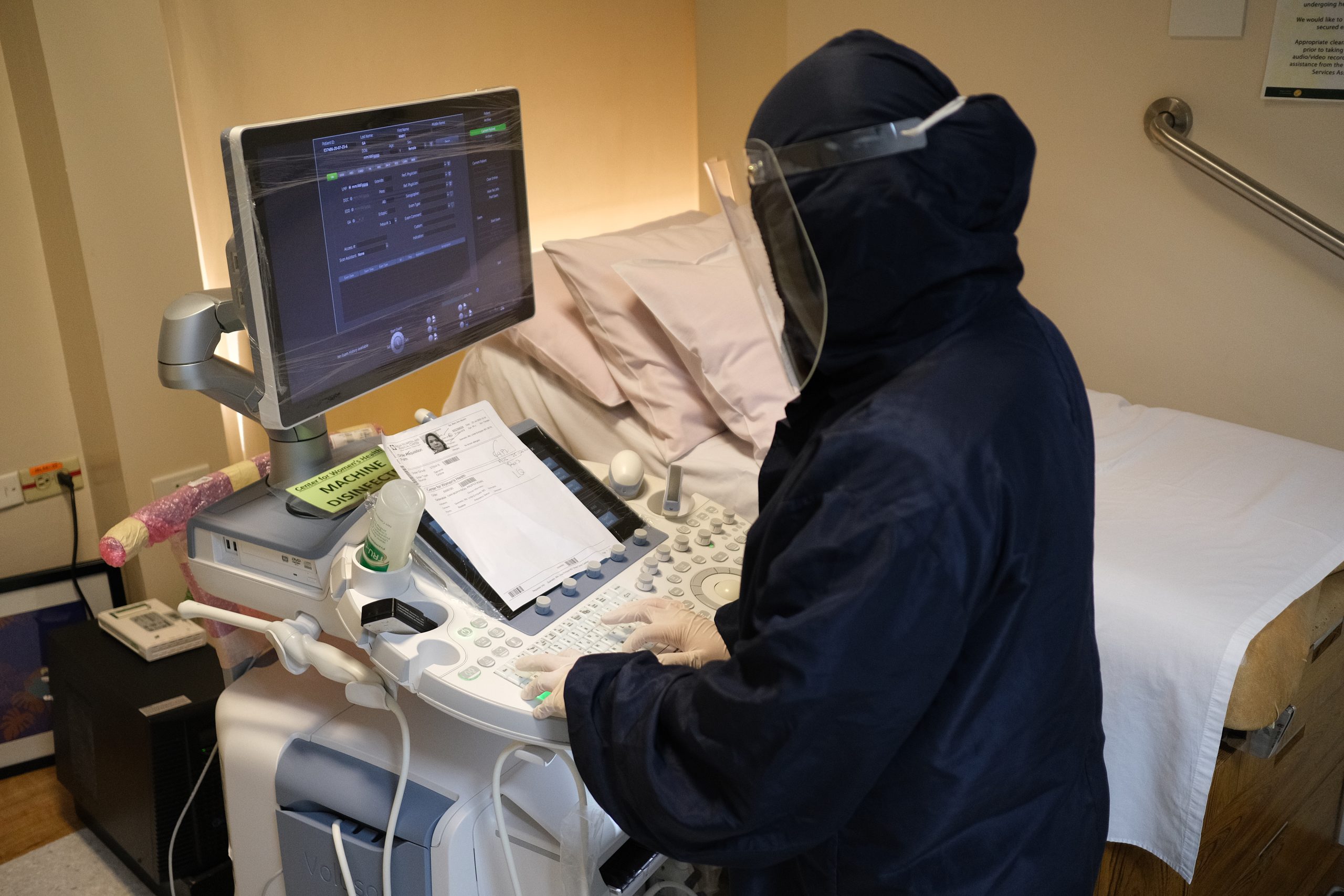
As a woman’s body develops with age, so does her healthcare needs. At the Asian Hospital and Medical Center, we are committed to addressing the unique healthcare needs of women of all ages and seasons in life. Our Center for Women’s Health is open to provide both non-invasive and minimally invasive ultrasound procedures and fetal surveillance to both in and out patients.
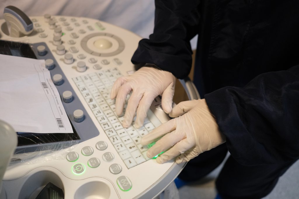
With a team of compassionate and experienced OB-GYN sonologists, perinatologists and clinical assistants, our unit delivers topnotch ultrasound service partnered with superior-quality care driven by our core values of credibility, honesty and sensitivity.
The Center for Women’s Health conducts 2D and 3D gynaecological ultrasounds to carefully assess the pelvis and accurately diagnose structural or functional disorders affecting the reproductive organs. Available to all women, our diagnostic procedures include:
- 2D ultrasound – This traditional form of ultrasound uses no-radiation, high-frequency sound waves in a single plane to capture live, two-dimensional images of the pelvic organs and transmit these images on a screen in real time.
- Transvaginal ultrasound (endovaginal ultrasound) – Meaning “through the vagina”, this is a type of internal examination in which the transducer is inserted into the vagina, unlike standard abdominal and pelvic ultrasounds where the transducer is pressed against the skin. A transvaginal ultrasound provides a clearer view of the ovaries, fallopian tubes, uterus and cervix.
- Transrectal ultrasound – Similar to a transvaginal ultrasound, a transrectal ultrasound offers internal examination of the pelvic organs. However, this test is inserted into the rectum and may be used to detect abnormalities in the rectal region and the prostate gland (in men).
- Follicle monitoring (follicle tracking) – This is a transvaginal ultrasound test used to determine when a woman is ovulating, specifically when a follicle matures and releases an egg. It is commonly done to help couples identify their fertile window and increase their chances for conception.
- Hysterosalpingosonogram (HSSG) – This refers to a sonographic scan that assesses any problems in the uterus, endometrium and fallopian tubes. Here, a special dye or saline solution is first injected through the cervix and into the uterus, allowing for clearer visualization of the organs during the transvaginal ultrasound.
- Saline infusion sonohysterography (SISH) – This sonographic scan offers a fairly rapid and highly accurate method of assessing the uterus and endometrium. Using a catheter, sterile saline solution is injected into the uterus during a transvaginal scan to better visualize and evaluate the uterus and its lining.
- 3D gynecologic ultrasound – A 3D ultrasound merges together a series of 2D images taken from different angles, providing a more detailed visualization of the pelvic organs. Aside from being used to assess a baby’s development during pregnancy, 3D ultrasounds are also helpful in diagnosing congenital anomalies, polyps and tumors, and uterine adhesions, and detecting intrauterine devices (IUDs).
The Center for Women’s health also offers ultrasound services to deliver accurate and efficient maternal and fetal healthcare, as folliows:
- 3D/4D Ultrasound – Both scans produce clearer 3D images of a developing baby compared to a standard 2D scan. 3D ultrasound scans produce still images that can be used to check that the baby is developing normally. 4D scans (with time as the 4th dimension) show moving images of the baby. This procedure is best done during the 26th to 31st week of pregnancy and comes with a package containing a CD and 2 colored pictures.
- OB Doppler velocimetry – This type of ultrasound measures the speed and direction of blood flow in the blood vessels. During pregnancy, a Doppler ultrasound is done to ensure that the blood flow from the uterus to the placenta is unrestricted. It also helps diagnose problems such as high-risk pregnancies due to preeclampsia, ectopic pregnancy or low amniotic fluid.
- Nonstress test – This is a non-invasive prenatal test done on the third trimester (28 weeks) to ensure that the fetal heart rate correlates with his or her movements. This routine procedure is named “nonstress” because no stress is applied on the fetus. No medications or special preparations are necessary as well.
- Congenital anomaly scan (morphology scan) – Typically done during the 18th to 22nd week of pregnancy, this test assesses the position of the placenta, the condition of the umbilical cord and the level of amniotic fluid. It is done to check the health, development and structural normalcy of the baby.
- Biophysical score – This prenatal test combines the nonstress test and fetal ultrasound to evaluate the baby’s heart rate, breathing, muscle tone, movements and amniotic fluid level. It is commonly done during the last trimester of pregnancy.
The Center for Women’s Health is located at the 4/F Main Hospital Building and is open from Monday to Saturday from 8:00am to 4:00pm.
-
 4th Floor, Main Building, Asian Hospital and Medical Center Metro Manila
-
